Structure Of Mammalian Eye
Hence it does not possess a perfect spherical shape. The fibrous tunic the vascular tunic and the nervous tunic.
 How Do We See Color We See Color Thanks To Specialized Receptors In Our Eyes Eye Facts Anatomy And Physiology Human Anatomy And Physiology
Light enters the eye by passing through the transparent cornea and aqueous humor.

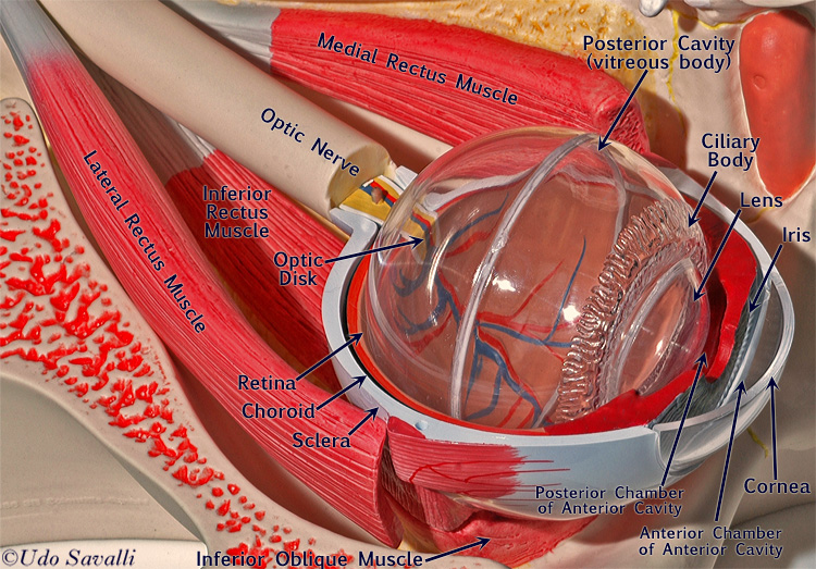
Structure of mammalian eye. The fibrous tunic the vascular tunic and the nervous tunic. The anterior segment is divided into two chambers. The basic function of these muscles is to provide different tensions and torques that further control the movement of the eye.
The structure of the mammalian eye has a laminar organization that can be divided into three main layers or tunics whose names reflect their basic functions. General structure of the mammalian eye. For this reason the developing eye has presented an invaluable system for studying the interactions among cells and more recently genes in specifying cell fate.
The fibrous tunic also known as the tunica fibrosa oculi is the outer layer of the eyeball consisting of the cornea and sclera. The fibrous tunic the vascular tunic and the nervous tunic. Structure of the Mammalian Eye.
The basic structure of the eye is similar in all mammals. Diagram of a scleral buckle. Anatomically the eye comprises two components fused into one.
As the animal was once living it deserves to be treated with respect. The front section anterior segment extends from the inside of the cornea to the front surface of the lens. External components include structures which can be seen on the exterior of the eye and internal components include structures present within.
Influence of the environment on the optical arrangements of animal eyes see text. General structure of the mammalian eye. The structure of the mammalian eye can be divided into three main layers or tunics whose names reflect their basic functions.
Functions of Mammalian Eye. The front anterior chamber extends from the cornea to the iris. Light rays enter the eye ball through the transparent cornea and are focussed by the lens on the retina.
To perform a dissection to understand the structure and function of the mammalian eye. The main parts of the human eye are the cornea iris pupil aqueous humor lens vitreous humor retina and optic nerve. They have the same parts and work in the same ways.
But the lens that performs the final focusing of light is found inside the eye behind the pupil. Specialized eyes of the chameleon Chamaeleo and the gecko Gekko. The _____is also known as the white of the eye.
The inverted image is corrected and a direct image is formed. The structure of the mammalian eye has a laminar organization that can be divided into three main layers or tunics whose names reflect their basic functions. There is a thick layer of fat between the eye and the orbital bones which act as shock absorber.
The _____ is the transparent front part of the eye. The lens is a complex structure. The fibrous tunic also known as the tunica fibrosa oculi is the outer layer of the eyeball consisting of the cornea and sclera.
An eye also consists of six muscles. These muscles move the eye in the orbit Each eye is about one inch in diameter and consists of three concentric layers- Sclera Choroid Retina 4. Mammalian vision is the process of mammals perceiving visible electromagnetic radiation analyzing it and forming subjective sensations on the basis of which the animals idea of the spatial structure of the external world is formed.
Birds and some reptiles have 3 eyelids. The sclera is essentially the continuation backward of the cornea the collagen fibres of the cornea being in. A set of six muscles attach the eye to the orbital bones.
Structure of the human eye. It has layers of soft tissue surrounding a firm nucleus. Mammals have two eyelids one upper and one lower eyelid of which the upper is more moveable.
Responsible for this process in mammals is the visual sensory system the foundations of which were formed at an early stage in the evolution of chordates. Three embryonic tissue sourcesthe neural ectoderm the surface ectoderm and the periocular mesenchymecontribute to the formation of the mammalian eye. An inverted image of the object is formed on the retina and this is conveyed to the visual centres of the optic lobes in the brain by the optic nerves.
It is filled with a fluid called the aqueous humor which nourishes the internal structures. It is made of an elastic capsule containing proteins and water which refract light at a constant rate just like the lenses used in glasses. The structure of the mammalian eye can be divided into three main layers or tunics whose names reflect their basic functions.
Visit an eye care unit or clinic with your friends and teacher to observe various charts displaying the anatomy of eye. The iris controls the size of the pupil which is the opening that allows light to enter the lens. Learn about this topic in these articles.
They can be closed to protect the eye from physical assault or from excess light. The Most Important Parts Of A Mammals Eye The Eyelids. Our eyelids are used in cleaning and protecting the eye.
The fibrous tunic the vascular tunic and the nervous tunic. The _____ is a gel-like substance that helps to keep the eyeball in its proper shape. It includes the medial rectus lateral rectus superior rectus inferior rectus inferior oblique and superior oblique.
 Human Eye Anatomy Parts Of The Eye Explained Eye Anatomy Anatomy Human Anatomy And Physiology
 Diagram Of Basic Parts Of The Eye Eye Anatomy Parts Of The Eye Diagram Of The Eye
 Visual System Sensory System Part 1 Sensory System Physiology Eye Anatomy
 Medical Textbook In The Net Eye Anatomy Eye Anatomy Medical Illustration Medical Anatomy
 Pin On Ophthalmology
 Eye And Ear Models Eye Anatomy Anatomy Models Medical Anatomy
 3d Human Anatomy Human Anatomy Atlas For Windows Desktop Human Anatomy And Physiology Human Anatomy Ciliary Muscle
 Human Eye Anatomy Eye Anatomy Anatomy Human Eye
 Cross Section Through The Human Eye Human Body Systems Human Anatomy And Physiology Body Systems
 Human Eye Anatomy Eye Anatomy Basic Anatomy And Physiology Human Anatomy And Physiology
The Human Eye Parts Of The Eyeball Human Eye Parts Of The Eye
 Structure Of The Human Eye
 Pin By Vaishnavitiws On Medical Map Chart Medical
 Vascular Supply Of Eye Anatomy Tendon Of Superior Rectus Muscle Veins Draining Scleral Venous Sinus Eye Anatomy Body Anatomy Human Anatomy And Physiology
 The Science Of Eye Health Exercises For Injuries Eye Anatomy Human Eye Diagram Eye Anatomy Diagram
 Pin By Brooke Bourgeois On School Stuff Human Anatomy And Physiology Medical Anatomy Eye Anatomy
 This Video Summarizes The Eye It Focuses On How Structures Work Together To Refract Light In Order To Focus On The Reti Eye Anatomy Human Eye Parts Of The Eye
 Ency123 Learn Create Have Fun How Does The Human Eye Work Neusholte Bril Eczeem
Post a Comment for "Structure Of Mammalian Eye"