2 Functions Of The Sclera
Structures in the Eye Functions 1. The sclera is a tough fibrous opaque tissue.
 Diagram Of Basic Parts Of The Eye Eye Anatomy Parts Of The Eye Diagram Of The Eye
Sclera is thickest posteriorly1mm.

2 functions of the sclera. The sclera is covered by the conjunctiva a. The primary function of the sclera is to protect the eye and maintain the shape of the eye ball. 2 the scleral epithelium is thicker than the corneal epithelium.
And 3 it has small basal cells with scanty cytoplasm. Its function is to provide strength and structure. 3 Functions of the Sclera 1.
The sclera is a dense connective tissue that accounts for five-sixths of the outer coat of the eyeball. The sclera is known as the white part of the eye. The sclera forms the entire visible white exterior of the eye the iris is the colored portion inside the anterior chamber of the eye.
Sclera Function The sclera along with the intraocular pressure IOP of the eye maintains the shape of the eyeball. 15 Both are soft connective tissues designed to provide structural integrity of the globe and to protect the inner components of the eye from phy si-. It is continuous with the dura mater and the cornea and maintains the shape of the globe offering resistance to internal and external forces and provides an attachment for the extraocular muscle insertions.
The sclera is the part of the eye commonly known as the white It forms the supporting wall of the eyeball and is continuous with the clear cornea. NETHRADHAMA SCHOOL OF OPTO 2. Lets Review Activity 2.
It is made of three regions. Six muscles attached to your. The sclera is the white part of your eye.
List two functions of the sclera. And the sclera Fig. Its a tough protective covering and the muscles that control eye movement are connected to it.
Withstands the considerable expansive force generated by the intraocular. The white part of the eye is called Sclera. While we can only see the visible portion of the sclera it.
The lamina fusca is the innermost region of elastic fibers. Without moving your head look up. Rods are responsible for night and peripheral side vision.
The episclera the sclera proper and the lamina fusca. What is the function of the optic nerve. Most of your eyeball is filled with a gel-like fluid called the vitreous humor.
Add your answer and earn points. The tough fibrous nature of the sclera also protects the eye from serious damage such as laceration or rupture from external trauma. Choroid 1 See answer yangedmark is waiting for your help.
1 2 Scleral thickness at the entry point of the optic nerve in the sheep is 10 to 12 mm. List two other functions of the cornea. Its whole outer surface is covered by tenons capsule and also by the bulbar conjunctiva in the anterior part Its inner surface lies in contact with the choroid with a potential suprachoroidal space in bertween.
Give the functions of the following parts of your eye. It along with the intraocular pressure IOP of the eye helps maintain the shape of your eyeball. 1 the collagen fibrils of the sclera are irregularly arranged.
Cones are responsible for sharp detailed central vision and color vision and are clustered mainly in the macula. The sclera differs from the cornea in three basic ways. There are two main types of photoreceptors.
The sclera is tough and fibrous protecting the interior components of the eye from injury and makes up the exterior coating of the eye. The following diagram shows the sclera. List two functions of the Sclera Gives the eye shape and helps protect delicate inner parts The cornea covers and helps protect the eye list two other functions of the cornea.
Introduction The sclera forms the posterior opaque 56 part of the external fibrous tunic of the eyeball. Examine the fat and muscle surrounding the eyeball. The sclera forms the posterior five-sixths of the connective tissue coat of the globe.
Maintains shape of the globe 2. The cornea covers and helps protect the eye. The sclera also protects the inner structures of your eye from trauma.
The sclera remarkable for its strength and firmness the word sclera is derived from the Greek sklera mannix which means hard membrane protects intraocular components from trauma light and mechanical displacement. It also provides protection to the interior parts of the eye.
 Baghali What Is The Human Eye Human Eye Eye Anatomy Eyes
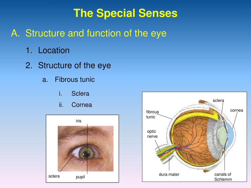 Ppt The Special Senses Powerpoint Presentation Free Download Id 873900
 The Eye Is A Sensory Organ That Perceives Light Transforms It Into An Electrical Signal And Sends It To Th Human Anatomy And Physiology Human Eye Eye Anatomy
 This Activity Is A Good Introduction To The Anatomy And Functions Of The Eye Students Will Identify Health And Physical Education Parts Of The Eye Fun Science
Eye Anatomy And Eye Diagram Iris Pharma Eye Anatomy Eye Anatomy Diagram Anatomy
 The Eye Diagram And Functions Functions Of The Human Eye Anatomy Body System Human Eye Diagram Eye Anatomy Diagram Of The Eye
 Wmu Psychology Department Lisa Baker Cow Eyes Anatomy Bones Anatomy
 Sclera White Of The Eye Medical Anatomy Eye Structure Human Eye Diagram
 Prevent Vision Loss And Eye Diseases With This Drink Healthzone Tips Eye Health Benefits Of Drinking Coffee Crohns Disease
 Parts Of An Eye Worksheet Education Com Parts Of An Eye 6th Grade Science Life Science
 Visual Perceptual Skills Keys To Learning Eye Anatomy Diagram Of The Eye Parts Of The Eye
 There Are Three Layers Or Tunics Of The Eyeball The Fibrous Layer Is The Outermost Layer That Contains The Medical Knowledge Medical School Studying Midterm

 The Canal Of Schlemm Sinus Venosus Sclerae Is A Passageway Inferior To The Aqueous Humor Of The Eye This Canal Drains Aqueous Eye Anatomy Anatomy Human Eye
 Cow Eye Dissection Cow Eyes Dissection Parts Of The Eye
 Alat Kesehatan Dan Laboratorium Gejala Kelainan Pada Mata Throat Anatomy Arteries And Veins Body Anatomy
 Anatomy Of The Eye Coloring Human Body Printables Human Body Worksheets Parts Of The Eye
 A Diagram Showing The Parts Of The Eye Including The Cornea Lens Iris And Ciliary Muscle At The Front Of The Eye Facts Human Eye Diagram Diagram Of The Eye
 Vision Parts Of The Eye 1 The Sclera 2 The Cornea 3 Conjunctiva 4 The Choroid Coat 5 The Ciliary Body 6 The Iris 7 The Turquoise Eyes Eye Drawing Purple Eyes
Post a Comment for "2 Functions Of The Sclera"